
Researchers from the Department of Clinical Sciences, Swedish University of Agricultural Sciences, Uppsala, Sweden (SLU) and the Department of Surgery, Blå Stjärnans Djursjukhus, Gothenburg, Sweden; Michael Pujari-Palmer, Odd Viking Höglund, Gustaf Svensson, and Tobias Per Otto Lundin, recently published a paper which examines the fixation capabilities of OsSticTM technology in a canine ex vivo tarsal arthrodesis test model.
Arthrodesis of the tarsal joint has a high incidence of postoperative complications and intraoperative difficulties. An adhesive that facilitates implant placement and joint alignment, as well as strengthening the internal fixation, could potentially be a useful complementary clinical tool to reduce the risk of complications in these high-risk procedures.
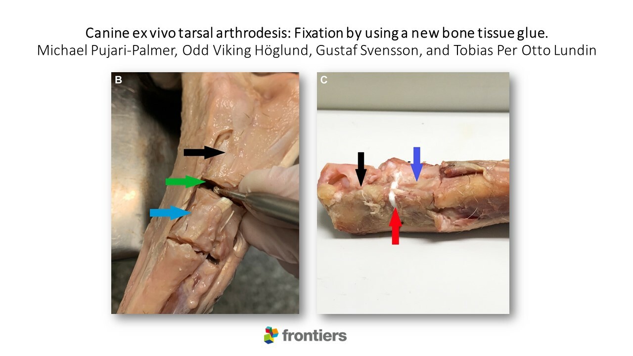
In this experimental ex vivo study, a new bone glue – OsSticTM technology, was evaluated as a subchondral bone adhesive. This glue is composed of materials that are present in the body; calcium and silicate, and the amino acid phosphoserine.
The biomechanical model the research group developed was sensitive enough to measure differences in fixation strengths between different glue formulations. The average fixation strength was 60–100 N/cm2, which should be strong enough to support short-term load bearing in medium sized canines (20 kg).
This demonstrates that this new resorbable glue – OsSticTM, can potentially contribute to stability at arthrodesis surgery, acting as a complement to today’s standard fixation, metal implants.
Read the full publication here.